An ECG is one of the most important and widely used diagnostic tools in cardiology. Simple, painless, and completed in minutes, it provides your cardiologist with a direct window into the electrical activity of your heart — revealing information that cannot be obtained through physical examination alone. At Vikram Heart Care Centre, ECG is available as part of our comprehensive cardiac diagnostic services, interpreted by Dr Kushaal Vikram with specialist cardiology expertise.
What Is an ECG?
An electrocardiogram — commonly referred to as an ECG or EKG — is a non-invasive test that records the electrical signals produced by the heart with each beat. Every time the heart contracts, an electrical impulse travels through the heart muscle in a precise sequence, triggering the chambers to pump blood in the correct order and rhythm.
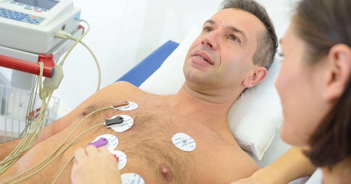
Small electrodes are attached to the skin of the chest, arms, and legs. These electrodes detect the electrical activity of the heart and transmit it to the ECG machine, which produces a graphical record — the characteristic waveform trace — that represents each phase of the cardiac cycle.
This trace is then analysed by a cardiologist to assess the heart’s rate, rhythm, and electrical conduction, and to identify signs of damage, strain, or abnormality.
What Does an ECG Measure?
A standard 12-lead ECG provides 12 different views of the heart’s electrical activity by recording from multiple electrode positions simultaneously.
Key information obtained includes:
- Heart rate — whether the heart is beating too fast (tachycardia), too slowly (bradycardia), or within the normal range
- Heart rhythm — whether the rhythm is regular or irregular, and whether it is a normal sinus rhythm or an arrhythmia
- Conduction — how electrical signals travel through the heart; delays or blocks in the conduction system are visible on the trace
- Chamber size — enlargement of the heart chambers produces characteristic changes in the waveform
- Signs of ischaemia — reduced blood flow to the heart muscle (from coronary artery disease) produces specific ECG changes
- Signs of infarction — evidence of a previous or ongoing heart attack appears as characteristic patterns on the trace
- Electrolyte abnormalities — dangerous levels of potassium, calcium, and other electrolytes affect cardiac electrical activity and are visible on ECG
- Drug effects — certain medications affect the ECG and monitoring is required during treatment
What Conditions Can an ECG Detect?
An ECG is valuable in diagnosing or providing evidence of a wide range of cardiac conditions:
Arrhythmias and Rhythm Disorders
- Atrial Fibrillation (AF) — the most common sustained cardiac arrhythmia; the ECG shows an irregularly irregular rhythm with no distinct P waves
- Atrial Flutter
- Supraventricular Tachycardia (SVT)
- Ventricular Tachycardia (VT) and Ventricular Fibrillation (VF)
- Heart Block (1st, 2nd, and 3rd degree) — delays or complete interruption of conduction between the upper and lower chambers
- Bundle Branch Block (LBBB and RBBB) — abnormal conduction through the ventricular walls
- Sick Sinus Syndrome
- Wolff-Parkinson-White Syndrome (WPW) — a condition involving an extra electrical pathway causing episodes of rapid heart rate
Coronary Artery Disease and Ischaemia
- ST elevation myocardial infarction (STEMI) — a classic heart attack pattern requiring emergency treatment
- Non-ST elevation myocardial infarction (NSTEMI)
- Stable angina — ST depression or T wave changes during exercise or stress
- Silent ischaemia — ECG changes in the absence of symptoms
Structural Heart Disease
- Left ventricular hypertrophy — thickening of the main pumping chamber, commonly from hypertension
- Right ventricular hypertrophy — from pulmonary hypertension or chronic lung disease
- Atrial enlargement — associated with valve disease, heart failure, and atrial fibrillation
Other Conditions
- Pericarditis — inflammation of the sac around the heart
- Pulmonary embolism — a blood clot in the lungs produces characteristic ECG changes
- Electrolyte disturbances — hyperkalaemia, hypokalaemia, hypercalcaemia
- Channelopathies — inherited conditions affecting cardiac ion channels, including Long QT Syndrome and Brugada Syndrome
Types of ECG
Resting 12-Lead ECG The standard ECG performed at rest, lying still. Takes approximately 5 to 10 minutes from electrode placement to completed recording. Provides a snapshot of cardiac electrical activity at that moment in time.
Exercise ECG (Treadmill Test / TMT) An ECG recorded continuously while the patient walks on a treadmill at progressively increasing speeds and inclines. Used to detect ischaemia that only becomes apparent when the heart is under the demand of exercise. Covered in detail in our TMT service page.
Holter Monitor (Ambulatory ECG) A portable ECG device worn continuously for 24 to 48 hours (or longer) during normal daily activities. Invaluable for capturing arrhythmias that occur intermittently and are not present during a brief resting ECG. Particularly useful for investigating palpitations, dizziness, or unexplained syncope.
Event Monitor A longer-term recording device worn for weeks to months that the patient activates when symptoms occur. Used when symptoms are very infrequent and unlikely to be captured by a standard Holter recording.
Who Should Have an ECG?
An ECG is recommended in a wide range of clinical situations. Your cardiologist may request one if you are experiencing symptoms or as part of a routine assessment.
Symptoms that warrant an ECG:
- Chest pain or chest tightness
- Palpitations — awareness of the heart beating, fluttering, or pounding
- Shortness of breath at rest or on exertion
- Dizziness, light-headedness, or fainting (syncope)
- Fatigue or exercise intolerance unexplained by other causes
- Swelling of the legs or ankles
Situations requiring an ECG as part of assessment:
- Pre-operative cardiac screening before surgery
- Newly diagnosed hypertension
- Diabetes mellitus — annual cardiac monitoring
- Starting or monitoring medications known to affect the heart
- Family history of sudden cardiac death or inherited cardiac conditions
- Routine cardiac health screening in adults over 40
- Following a stroke or transient ischaemic attack (TIA) to check for atrial fibrillation
- Athletes undergoing pre-participation cardiac screening
The Procedure: What to Expect
An ECG is entirely painless and non-invasive. There is no electrical current delivered to the body — the electrodes only record signals, they do not transmit them.
What happens during an ECG:
- You will be asked to lie flat on an examination table
- Small sticky electrode patches are attached to specific positions on your chest, arms, and legs
- You will be asked to lie still and breathe normally for approximately 10 seconds while the recording is taken
- The electrodes are removed and the recording is complete
- There is no discomfort, no needles, and no radiation
The entire procedure from preparation to completion takes approximately 5 to 10 minutes. Results are interpreted by Dr Kushaal Vikram and discussed with you at the same appointment or shortly thereafter.
Limitations of a Resting ECG
While enormously valuable, a resting ECG has important limitations that are worth understanding:
- A normal resting ECG does not rule out significant coronary artery disease — blockages in the coronary arteries may only produce ECG changes during exercise or stress
- Intermittent arrhythmias may not be present at the time of recording; a Holter monitor may be required
- An ECG provides electrical information only — structural abnormalities such as valve disease are better evaluated by echocardiography
- An ECG is one piece of a diagnostic puzzle; findings must always be interpreted in the context of clinical history, symptoms, and other investigations
Frequently Asked Questions
Q: Is an ECG safe?
An ECG is completely safe. No electricity is delivered to your body — the electrodes simply sense and record the electrical signals your heart already produces naturally. There is no radiation involved. It is safe for pregnant women, children, and elderly patients.
Q: Can I eat or drink before an ECG?
Yes. Unlike some other cardiac tests, a resting ECG requires no special preparation. You can eat, drink, and take your usual medications normally before the test.
Q: My ECG was reported as abnormal. Does that mean I have heart disease?
Not necessarily. Some ECG findings that are reported as abnormal variants are not clinically significant — they simply reflect normal variation in electrical patterns between individuals. Others do indicate a condition requiring further investigation. An abnormal ECG result should always be discussed with a cardiologist who can interpret it in the context of your symptoms, history, and overall clinical picture.
Q: I had an ECG during a health check-up and it was normal. Can I still have a heart problem?
Yes. A normal resting ECG is reassuring but does not exclude all cardiac conditions. Coronary artery disease, for example, may only produce ECG changes during exercise. Structural problems such as valve disease require echocardiography to detect. If you have cardiac symptoms despite a normal resting ECG, further investigation is warranted.
Q: How often should I have an ECG?
There is no universal recommendation for routine ECG screening in the general population. However, a baseline ECG is reasonable for adults over 40, particularly those with risk factors such as hypertension, diabetes, a family history of heart disease, or significant smoking history. Your cardiologist will advise on the appropriate frequency for your individual situation.
Q: My child has been referred for an ECG. Should I be worried?
ECG referrals in children are common and do not always indicate a serious problem. They are frequently requested to investigate palpitations, fainting episodes, chest pain, or to screen children with a family history of inherited cardiac conditions. The test is entirely safe and painless for children.
Q: Is ECG available in Patna?
Yes. ECG is available at Vikram Heart Care Centre, Dr K K Kantha Memorial Hospital, 21 B/3, Patliputra Colony, Patna.
Book a Consultation
If you are experiencing chest pain, palpitations, breathlessness, or dizziness, or if you require a cardiac assessment for any reason, an ECG is often the first and most important step.
📞 +91 893 583 4142 | +91 886 400 4584
📍 21 B/3, Patliputra Colony, Near Pataliputra Golambar, Opp. UNICEF Building, Patna 800 013
🌐 Book online at kanthahospital.com