Coronary angiography is the gold standard diagnostic procedure for evaluating the coronary arteries — the blood vessels that supply the heart muscle with oxygen-rich blood. When non-invasive tests such as an ECG, echocardiogram, or treadmill test suggest the possibility of coronary artery disease, angiography provides the definitive answer: a direct, high-resolution image of the coronary circulation that reveals exactly where blockages exist, how severe they are, and what treatment is needed.
At Vikram Heart Care Centre, coronary angiography is performed by Dr Kushaal Vikram in a fully equipped cardiac catheterisation laboratory, with the expertise and infrastructure to move seamlessly from diagnosis to intervention when required.
What Is Coronary Angiography?
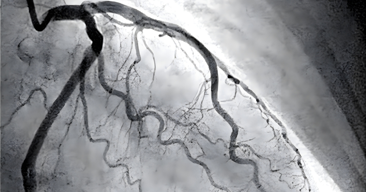
Coronary angiography — also called cardiac catheterisation or a coronary angiogram — is a minimally invasive procedure in which a thin, flexible tube called a catheter is guided through the blood vessels to the coronary arteries. A contrast dye is injected through the catheter, and X-ray imaging captures the dye as it flows through the arteries, making the vessels and any blockages or narrowings clearly visible on a live fluoroscopic screen.
The procedure is performed in a specialised room called a cardiac catheterisation laboratory (cath lab), equipped with high-resolution X-ray imaging systems, monitoring equipment, and emergency facilities.
Why Is Coronary Angiography Performed?
Angiography is recommended when there is clinical suspicion of significant coronary artery disease, or when non-invasive tests have produced inconclusive or concerning results.
Common reasons for referral include:
- Chest pain (angina) that is new, worsening, or not responding to medical treatment
- A positive or inconclusive exercise stress test (TMT)
- Abnormal findings on a nuclear stress test or stress echocardiogram
- Suspected acute coronary syndrome — unstable angina or heart attack
- Evaluation before major cardiac surgery such as valve replacement or bypass surgery
- Investigation of unexplained heart failure or reduced heart function
- Evaluation of coronary artery anomalies detected on other imaging
- Recurrent symptoms after previous angioplasty, stenting, or bypass surgery
- High-risk occupations requiring definitive cardiac clearance
Understanding Coronary Artery Disease
The coronary arteries are three main vessels — the left anterior descending (LAD), the left circumflex (LCx), and the right coronary artery (RCA) — and their branches. These arteries supply different regions of the heart muscle.
Over time, fatty deposits called plaques can build up inside the arterial walls — a process called atherosclerosis. As plaques grow, they narrow the artery and restrict blood flow to the heart muscle. This reduced flow causes chest pain (angina) during exertion when the heart demands more oxygen than the narrowed artery can deliver.
If a plaque ruptures suddenly, a clot forms at the site, potentially blocking the artery completely. This is a heart attack — and it is a medical emergency requiring immediate intervention.
Coronary angiography maps this process with precision, identifying:
- Which arteries are affected
- The exact location and length of each narrowing
- The severity of stenosis (percentage of narrowing)
- Whether the narrowing is suitable for angioplasty, stenting, or bypass surgery
- The condition of the heart muscle supplied by the affected vessels
The Procedure: What to Expect
Before the Procedure
- You will be asked to fast for 4 to 6 hours before the procedure
- Blood tests, an ECG, and a review of your medications will be performed
- Medications that may need to be temporarily stopped include blood thinners and diabetes medications — your cardiologist will advise specifically
- You will be asked about allergies, particularly to contrast dye or iodine
- An intravenous (IV) line will be placed for medications and fluids
- The access site — usually the wrist (radial artery) or groin (femoral artery) — will be cleaned and numbed with local anaesthetic
During the Procedure
- You will lie on a padded X-ray table in the cath lab
- A small incision is made at the access site and a sheath is inserted into the artery
- The catheter is guided through the artery to the heart under continuous X-ray guidance — you will not feel this as arteries have no pain receptors
- Contrast dye is injected through the catheter into each coronary artery in turn
- X-ray images are captured as the dye flows through the vessels, clearly showing the coronary anatomy and any blockages
- When the dye is injected you may feel a brief warm or flushing sensation — this is normal and passes quickly
- The entire procedure typically takes 30 to 60 minutes
- If a significant blockage is found, your cardiologist may proceed directly to angioplasty and stenting in the same session (ad hoc PCI), or plan a separate procedure depending on the findings and your overall condition
After the Procedure
- The catheter and sheath are removed and pressure is applied to the access site to stop bleeding
- For radial (wrist) access, a compression band is applied for 2 to 4 hours
- For femoral (groin) access, you will need to lie flat for several hours
- Most patients are observed for 4 to 6 hours and can go home the same day for a diagnostic-only procedure
- You will need someone to drive you home
- The access site should be kept dry and monitored for bleeding or swelling
- Normal activities can typically resume within 24 to 48 hours
Radial vs Femoral Access
Modern coronary angiography is most commonly performed via the radial artery at the wrist, which has become the preferred approach at most centres due to:
- Lower risk of bleeding complications
- No requirement for prolonged bed rest after the procedure
- Greater patient comfort
- Earlier discharge and return to normal activity
The femoral (groin) approach may be preferred in certain anatomical or clinical situations where radial access is not suitable. Your cardiologist will choose the most appropriate approach for your individual case.
Understanding Your Results
After the procedure, your cardiologist will review the images and discuss the findings with you. Results broadly fall into the following categories:
Normal Coronary Arteries No significant narrowings are identified. This is reassuring and means that your symptoms are unlikely to be caused by obstructive coronary artery disease. Further investigation for other causes will be discussed.
Non-Obstructive Coronary Artery Disease Some plaque or wall irregularities are present but do not cause significant narrowing. Medical management with risk factor modification and medications is typically recommended.
Obstructive Coronary Artery Disease One or more significant narrowings are identified. Treatment options depend on the number, location, and severity of blockages:
- Single vessel disease — angioplasty and stenting (PCI) is usually the preferred treatment
- Two vessel disease — PCI or bypass surgery depending on the complexity of the lesions
- Three vessel disease or left main disease — bypass surgery (CABG) is often recommended, particularly in patients with diabetes or reduced heart function; a multidisciplinary heart team discussion guides the decision
Risks and Complications
Coronary angiography is a safe and routinely performed procedure. Serious complications are uncommon but exist and should be understood before consenting to the test.
Minor complications (more common):
- Bruising or haematoma at the access site
- Allergic reaction to contrast dye — mild reactions (rash, itching) are managed with medications; severe anaphylaxis is very rare
- Temporary kidney stress from contrast dye — patients with pre-existing kidney disease require special precautions and hydration
- Brief irregular heartbeat during catheter manipulation — usually self-terminating
- Vasovagal reaction — fainting or drop in blood pressure during or after the procedure
Major complications (uncommon):
- Serious bleeding requiring intervention
- Arterial injury at the access site
- Stroke — very rare
- Heart attack triggered by the procedure — very rare
- Contrast-induced nephropathy in patients with impaired kidney function
Your cardiologist will discuss these risks in detail and weigh them against the benefit of the diagnostic information the procedure provides.
Contrast Dye and Kidney Function
The contrast dye used in angiography is filtered by the kidneys. In patients with pre-existing kidney disease, diabetes, or dehydration, the dye can temporarily worsen kidney function — a condition called contrast-induced nephropathy (CIN).
Precautions taken to minimise this risk include:
- Pre-procedure kidney function blood tests
- Adequate hydration before and after the procedure
- Using the minimum effective volume of contrast dye
- Temporary withholding of certain medications such as metformin
- Post-procedure monitoring of kidney function
In patients with significantly impaired kidney function, the risks and benefits of contrast angiography are carefully weighed, and alternatives such as CT angiography with low-dose contrast or cardiac MRI may be considered.
Coronary Angiography vs CT Coronary Angiography
CT coronary angiography (CTCA) is a non-invasive alternative that uses a CT scanner with intravenous contrast to image the coronary arteries. It is useful for ruling out coronary artery disease in lower-risk patients with atypical symptoms.
Key differences:
- CTCA is non-invasive and does not require arterial catheterisation
- CTCA involves radiation from the CT scanner; invasive angiography uses X-ray fluoroscopy
- Invasive angiography provides higher resolution images and allows immediate treatment if a blockage is found
- CTCA cannot be followed immediately by angioplasty or stenting
- Invasive angiography remains the definitive gold standard, particularly in higher-risk patients or those in whom intervention is anticipated
Your cardiologist will advise which test is most appropriate for your clinical situation.
Frequently Asked Questions
Q: Will I be awake during the angiography?
Yes. Coronary angiography is performed under local anaesthesia with sedation. You will be awake but relaxed and comfortable. The access site is numbed completely so you will not feel the catheter insertion. You may feel a warm flushing sensation when contrast dye is injected — this is normal and passes within seconds.
Q: Is the procedure painful?
Most patients find coronary angiography much less uncomfortable than they anticipated. The local anaesthetic effectively numbs the access site. You may feel some pressure during catheter manipulation but not pain. The brief warm flush from contrast injection is the most commonly reported sensation. Any significant discomfort should be reported to the cath lab team immediately.
Q: How long will I need to stay in hospital?
For a diagnostic angiography via the radial (wrist) approach, most patients go home the same day after 4 to 6 hours of observation. If angioplasty and stenting are performed in the same session, an overnight stay is usually recommended. Your cardiologist will advise based on your individual findings and clinical status.
Q: What if a blockage is found during the angiography?
If a significant blockage is identified, your cardiologist will discuss the findings with you and explain the treatment options. In many cases, angioplasty and stenting can be performed in the same session — this is called ad hoc PCI. In others, particularly with complex multi-vessel disease, a planned procedure or surgical referral may be recommended after a multidisciplinary team discussion.
Q: I am diabetic. Are there special precautions I need to take?
Yes. Patients with diabetes require specific precautions before and after angiography. Metformin is typically withheld for 48 hours around the procedure due to the risk of lactic acidosis in the presence of contrast dye. Kidney function is monitored closely. Your cardiologist and team will give you specific instructions regarding your diabetes medications before the procedure.
Q: Can I drive myself home after the procedure?
No. You should not drive yourself home after coronary angiography. The sedation given during the procedure affects your reactions and judgement, and the access site requires monitoring. Please arrange for a family member or companion to drive you home.
Q: Is coronary angiography available in Patna?
Yes. Coronary angiography is performed in the fully equipped cardiac catheterisation laboratory at Vikram Heart Care Centre, Dr K K Kantha Memorial Hospital, Patna, by Dr Kushaal Vikram.
Book a Consultation
If you have been experiencing chest pain, breathlessness, or have received an abnormal result on a cardiac screening test, a consultation with Dr Kushaal Vikram will help determine whether coronary angiography is the right next step for you.
📞 +91 893 583 4142 | +91 886 400 4584
📍 21 B/3, Patliputra Colony, Near Pataliputra Golambar, Opp. UNICEF Building, Patna 800 013
🌐 Book online at kanthahospital.com